
You might notice one testicle looks a little larger than the other, or a scrotum that seems fuller than it used to. Sometimes it is subtler than that: a dog who is intact and getting older may just look a bit “different” around the back end, and it is easy to assume it is normal ageing.
Testicular tumours in dogs can be like that. Many dogs show few obvious signs until a lump is found by chance during a bath, a groom, or a routine vet visit.1 When something does change, it is not always a dramatic “cancer” picture. Hormone-related effects, skin and coat changes, or a dog who is simply not quite themselves can be part of the story too.1, 2
What matters in practice is not trying to diagnose it at home, but knowing what is worth checking and when to book in. Testicular tumours are often treatable, particularly when they are found before they have spread, and there are some clear risk factors that can help you and your vet make sensible decisions.1
Causes and risk factors

There is rarely one single “cause” for a testicular tumour. Most are thought to arise from a mix of genetic predisposition and age-related change, and we do not always know why a particular dog develops one while another does not.1
Age and being intact
Testicular tumours are seen most often in older, intact male dogs. This does not mean younger dogs cannot be affected, but the odds rise with time, and the cancers that occur in dogs tend to be different in behaviour and age profile from those in humans.1, 5
Cryptorchidism (retained testicle)
If a dog has one or both testicles that never descended into the scrotum (cryptorchidism), the risk picture changes. Retained testicles are linked with a significantly higher risk of certain testicular tumours, and also a higher risk of testicular torsion, which is a painful emergency.2, 5
Breed patterns
Some breeds are reported more often in association with testicular tumours, including Boxers, German Shepherd Dogs and Weimaraners. This is best thought of as a tendency, not a guarantee, and it should not replace regular physical checks and veterinary exams.1
Signs you might notice at home

The most common “early sign” is simply a lump, firmness, or uneven size in one testicle, or general swelling of the scrotum.1 Some dogs are not bothered by it at all, which can make it easier to miss.
Depending on the tumour type, there can also be hormone-related signs. Some Sertoli cell tumours can produce oestrogen, leading to changes sometimes described as feminisation, symmetrical hair loss, skin darkening, enlarged mammary tissue, or reduced interest in mating behaviour. In more serious cases, hormone effects can suppress bone marrow function and contribute to lethargy or paler gums.1
Less specific signs like weight loss, decreased appetite, vomiting, or difficulty urinating or defecating can occur if disease is advanced or has spread, but these are not the usual starting point.1
A simple, calm check you can do
You do not need a “medical” exam at home, just a bit of familiarity with what is normal for your dog.
- When your dog is relaxed, gently feel the scrotum and note whether the testicles feel smooth and similar in size (minor differences can be normal, sudden change is not).1
- Look for swelling, redness, heat, or discomfort.
- For dogs who have only one testicle in the scrotum, treat that as a reason to discuss cryptorchidism with your vet, even if the dog seems well.2
If you find a lump or your dog seems sore, book a veterinary appointment rather than waiting to “see if it goes away”. Other conditions, including infection, inflammation, injury, or benign growths, can look similar from the outside.1, 3
How vets diagnose testicular tumours
A veterinary visit usually starts with a history and physical exam, including palpation of the scrotum and a general check of lymph nodes and abdomen.1, 3 If a dog is cryptorchid, the vet may recommend imaging to locate the retained testicle.2
Imaging and staging
Ultrasound is commonly used to assess the testicle and surrounding tissues, especially when the testicle is retained in the abdomen. If cancer is suspected, your vet may discuss “staging” tests, which can include blood and urine tests and chest X-rays, to check for any signs of spread and to plan anaesthesia and surgery safely.1, 4
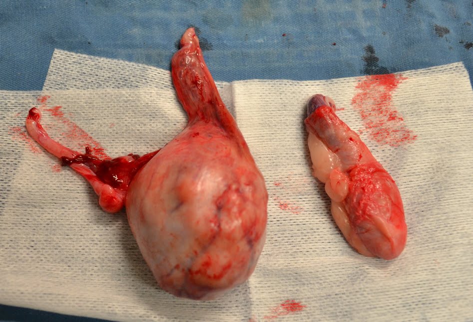
Confirming the tumour type
In many cases, the tumour type is confirmed after surgical removal (castration or removal of the affected testicle), when tissue is sent to a laboratory for histopathology. That report helps clarify whether the tumour is benign or malignant, and whether further treatment is likely to be helpful.1, 4
Treatment options and what they involve

For most dogs, surgery is the main treatment. Removing the testicles removes the tumour site and also allows the lab diagnosis that guides next steps.1 If the dog is cryptorchid, surgery is typically more involved because the retained testicle may be in the abdomen or inguinal region.2
If there is evidence that a tumour has spread, or if the tumour type is considered higher risk, your vet may recommend referral to a veterinary oncologist to discuss chemotherapy and, in selected cases, radiation therapy. These decisions depend on where disease is present, how quickly it is progressing, and what your dog is likely to tolerate well.1, 4
Aftercare and monitoring
Most dogs recover well after routine castration, but the plan should still be tailored. Your vet will advise on wound care, activity restriction, pain relief, and follow-up timing. If staging showed any concerns, or if histopathology suggests a meaningful risk of spread, monitoring may include rechecks and repeat imaging at intervals your vet considers appropriate.3
Prognosis and what “good outcome” really means
It is tempting to look for a single survival number, but prognosis is more useful when it is specific to tumour type and whether there is spread.1 Many canine testicular tumours behave as a local problem, and spread is uncommon for several common tumour types. That is one reason surgery alone can be curative in a large proportion of cases.1
Where tumours are malignant or have spread, outcomes are more variable. The best guide is the combination of histopathology results and staging tests, alongside your dog’s overall health and comfort. Your vet can help you weigh up treatment intensity, side effects, and the kind of day-to-day life your dog is likely to have with each option.4
Prevention and risk reduction

The most direct prevention is straightforward: neutering removes the organ where testicular tumours arise. For dogs not intended for breeding, it is a practical risk-reduction step, and it is particularly strongly recommended for dogs with cryptorchidism.2, 6
Timing is worth discussing. Desexing decisions also involve considerations like behaviour, orthopaedic development, and other health risks that vary by breed and individual dog. A good conversation with your vet can put cancer prevention in the wider context, rather than treating it as a single-issue decision.6
General health steps that still matter
There is no proven diet or supplement that “prevents” testicular cancer, but good general health makes diagnosis and treatment easier to manage. Keeping your dog at a healthy weight, staying current with routine veterinary checks, and avoiding unnecessary exposure to toxins are sensible foundations, even when the evidence is not specific to this one cancer.7
Living with a dog diagnosed with a testicular tumour
Once a diagnosis is on the table, the day-to-day goal becomes simple: keep your dog comfortable while you work through decisions and timelines. Most dogs cope well with the veterinary visits and the surgery itself, especially when pain relief and recovery plans are well managed.
It can help to keep notes on what you are seeing at home, including appetite, toileting, energy, interest in walks, and any licking or sensitivity around the groin. This gives your vet clearer information than memory alone, and it supports practical pain and comfort planning if treatment needs to be extended.
If you are considering chemotherapy or have been referred to an oncology service, ask about what side effects are common in dogs, what monitoring is needed, and what would count as a reason to pause or stop treatment. The aim is not to pursue “everything possible”, but to choose care that fits your dog and your household.
Final thoughts
Testicular tumours in dogs are often found in ordinary moments, a quick pat, a wash, a routine check-up, rather than through dramatic symptoms. That is why familiarity with your dog’s normal body, and a willingness to book a vet visit when something changes, can make such a difference.
For many dogs, treatment is as straightforward as surgical removal and a histopathology report to confirm what was there. For others, especially cryptorchid dogs or dogs with signs of spread, the pathway can be more involved. Either way, clear information and steady follow-up usually lead to better decisions than worry and waiting.
References
- VCA Animal Hospitals: Testicular Tumors
- Cornell University College of Veterinary Medicine: Cryptorchidism in dogs (retained testicle)
- MSD Veterinary Manual: Tumors of the Male Reproductive Tract in Small Animals
- PetMD: Testicular Tumors in Dogs
- Biology of Reproduction (Oxford Academic): Cryptorchidism and testicular cancer in the dog, unresolved questions and challenges
- RSPCA Australia: Desexing your pet
- RSPCA Knowledgebase: Health problems associated with obesity in dogs
- American College of Veterinary Surgeons: Castration (Neuter) in Dogs
- WSAVA: Global Nutrition Guidelines